O-scan MRI Scanner
The O-scan MRI scanner is characterised by high imaging quality with low installation and operating costs.
Clinical applications
High imaging quality at low cost
O-scan
APPLICATION
Ideal for radiology, orthopaedics, rheumatology and sports medicine departments.
FOR WHICH EXAMINATIONS?
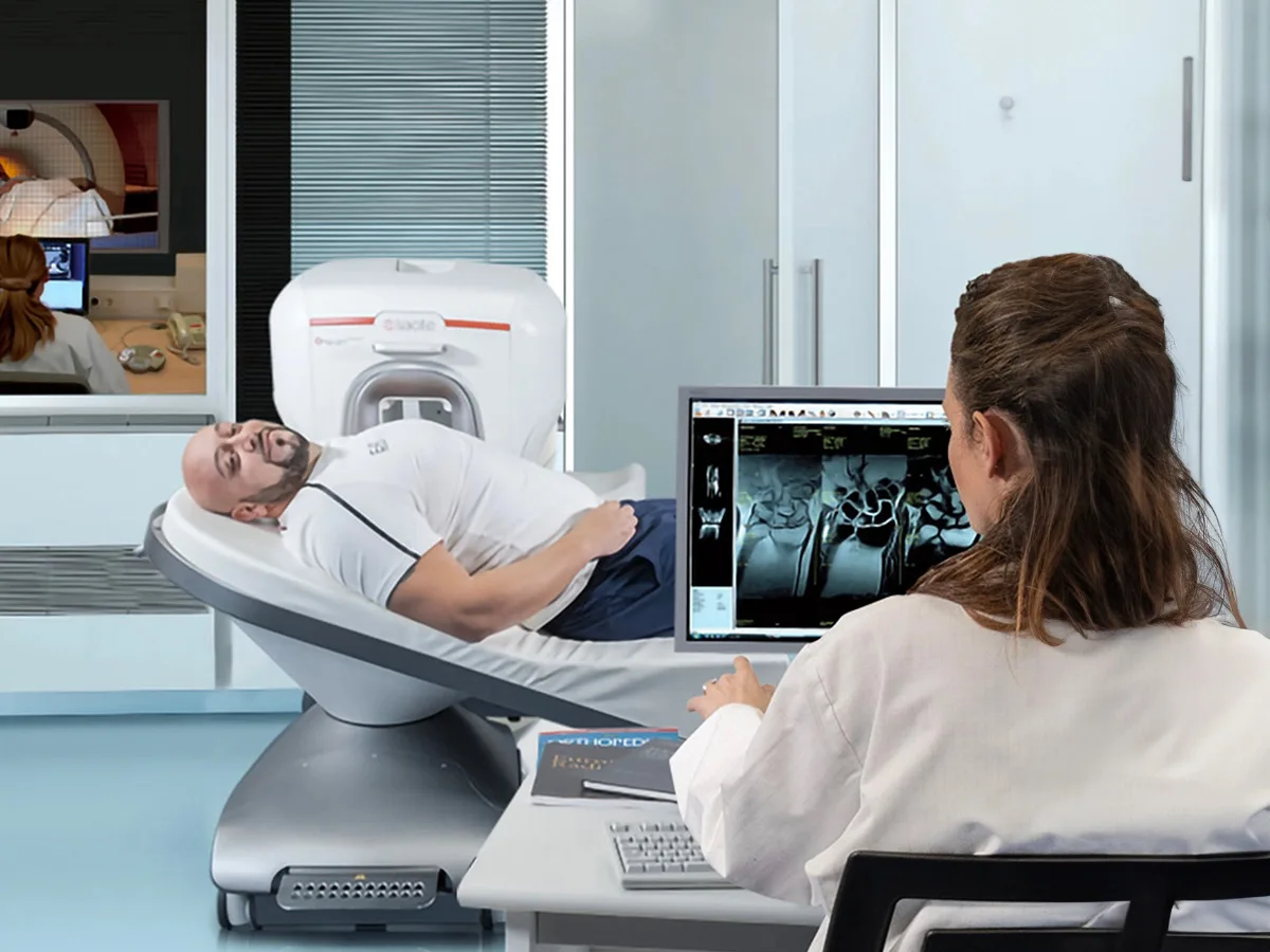
The O-scan magnetic resonance imaging system is a unique system dedicated to orthopaedic examinations (knee, ankle, foot, hand, wrist and elbow).
PURCHASE ECONOMICS
A very fast return on investment; the cost of an examination is just energy costs (about the same as an average electric kettle). Its low weight and small dimensions mean that the system can be installed in almost any room with an area of 9 m2
ESAOTE O-scan
The O-scan MRI scanner is characterised by high imaging quality with low installation and operating costs. No additional power supply is needed, and the O-scan can be placed on any type of floor. Moreover, it requires only a small area – the minimum required area is 9 m2.
The O-scan magnetic resonance imaging system was created with the idea of dedicated MSK-MRI applications in radiology, orthopaedics and sports medicine. It is an ideal 2nd MRI system in radiology practice, providing high image quality with minimal investment in space and finances. The O-scan is a cost-effective solution that relieves a large high-field system and an economically efficient solution for small orthopaedic practices.
The O-scan gantry features a self-centring coil system with automatic coil recognition, as well as a unique design that prevents RF interference.
Watch Video Materials about O-scan
General specifications
- A wide spectrum of orthopaedic examinations
- Maintenance-free permanent magnet
- Device housing designed to increase patient comfort and ease of positioning - no claustrophobic effect
- No additional power supply is needed - Single-phase power supply
- Energy consumption: < 1.5 KW
- O-scan can be placed on any type of floor
- System weight: 1,240 Kg
- RF shielding: Integrated internal shielding and a free-standing Faraday cage
- Minimum room dimensions with an external Faraday cage 3.20 m x 3.50 m for single-room installation
- Minimum room dimensions: a Shielding collar type Faraday cage, i.e. shields placed directly on the patient's limbs 2.80 m x 3.20 m for single-room installation
- Short scan time: 12-minute protocols (e.g. for knees)
- No claustrophobic effect.
SIMPLE OPERATION AND UNIQUE DESIGN
The device performs excellently in diagnostic orthopaedic practices and is an excellent alternative and complement to other MRI systems, relieving, for example, a high-field MRI scanner.
HIGH IMAGE QUALITY AND SHORT SCAN TIME
Standard throughput of 2 patients / hour including preparation procedures, fast 12-minute protocols (e.g. for knees).
LOW EXAMINATION COST
Only energy costs (about the same as an average electric kettle)
LOW ROOM ADAPTATION COSTS
An external Faraday cage or a Shielding collar type Faraday cage, i.e. shields placed directly on the patient's limbs; the single-phase power supply does not require power modifications as in the case of high-field MRI, air cooling (standard air conditioners)
Check the differences
Dedicated O-scan Magnetic Resonance Imaging vs High-Field Magnetic Resonance Imaging
- single-phase power supply
- no need to interfere with the existing installation
- does not require power modifications as in the case of high-field MRI
- low examination cost - only energy costs (about the same as an average electric kettle)
- low service costs - only calibration costs
- low room adaptation costs - external Faraday cage
- The need to create a separate 3-phase power supply with 100A protection
- the need to replace (replenish) the cooling agent
- electromagnet at specific time intervals - high costs
- high service costs
- high room adaptation costs (the need to build a Faraday cage and specialised air conditioning during room adaptation)
eXP Technology
Check the technologies that can be found in the O-scan
- True Motion – enables real-time imaging of moving joints. The entire sequence can be saved in the popular .avi format, so the film can be played on almost any device.
- MAR Metal Artifact Reduction – software that enables the reduction of image artefacts originating from metal implants.
- 3D Viewer – An advanced viewer for 3D images created through acquisition using isotropic 3D sequences.
- 2D/3D Speed Up - Software that, using mathematical algorithms, allows for a reduction in image acquisition and reconstruction time, up to 30% in 2D mode and as much as 50% in 3D mode, compared to the standard imaging mode.
- Isotropic 3D – advanced imaging of three-dimensional isotropic sequences, which makes it possible to combine Speed Up technology, enabling fast acquisition of high-resolution 3D images, with advanced 3D image reconstruction techniques – MPR. During a single acquisition, you obtain the highest quality image in all three planes.
- 3D Sharc – A three-dimensional sequence dedicated to high-resolution imaging of cartilage tissue.
Check the purchasing options for the device
See also
Magnifico VET MRI System
Magnetic resonance imaging - the new Esaote Magnifico offers revolutionary MRI solutions that increase diagnostic accuracy. Dedicated to veterinary medicine
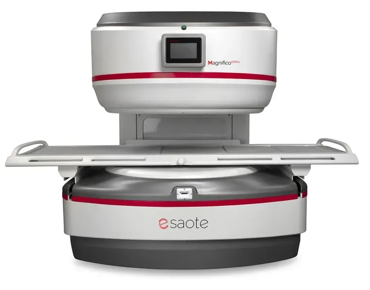
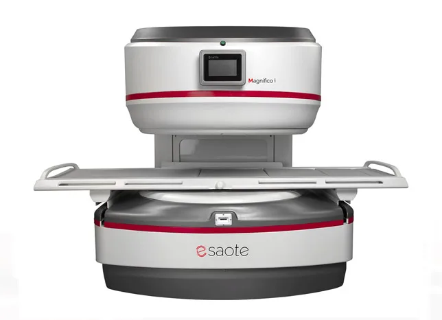
Magnifico Open MRI System
Magnifico Open is a next-generation open MRI system that combines the latest imaging technology with a permanent magnet for low operating costs, now powered by the e-SPADES fully integrated AI solution.
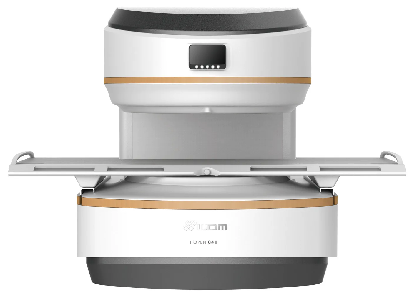
I_Open 0.36T/0.4T MRI Scanner
The i_Open 0.36T/0.4T MRI scanner is a modern magnetic resonance imaging system with a permanent magnet generating a field of 0.36T or 0.4T.
Request a quote for O-scan MRI Scanner
You will receive a quote, specifications and an offer of a free demonstration. We usually reply the same business day.
- Individual quote
- Free demonstration at your facility
- Support from our in-house service department